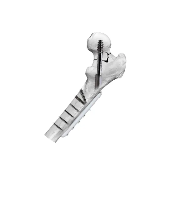
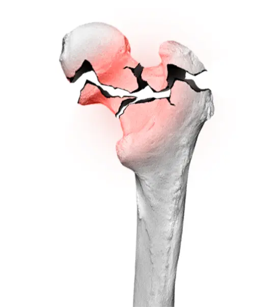
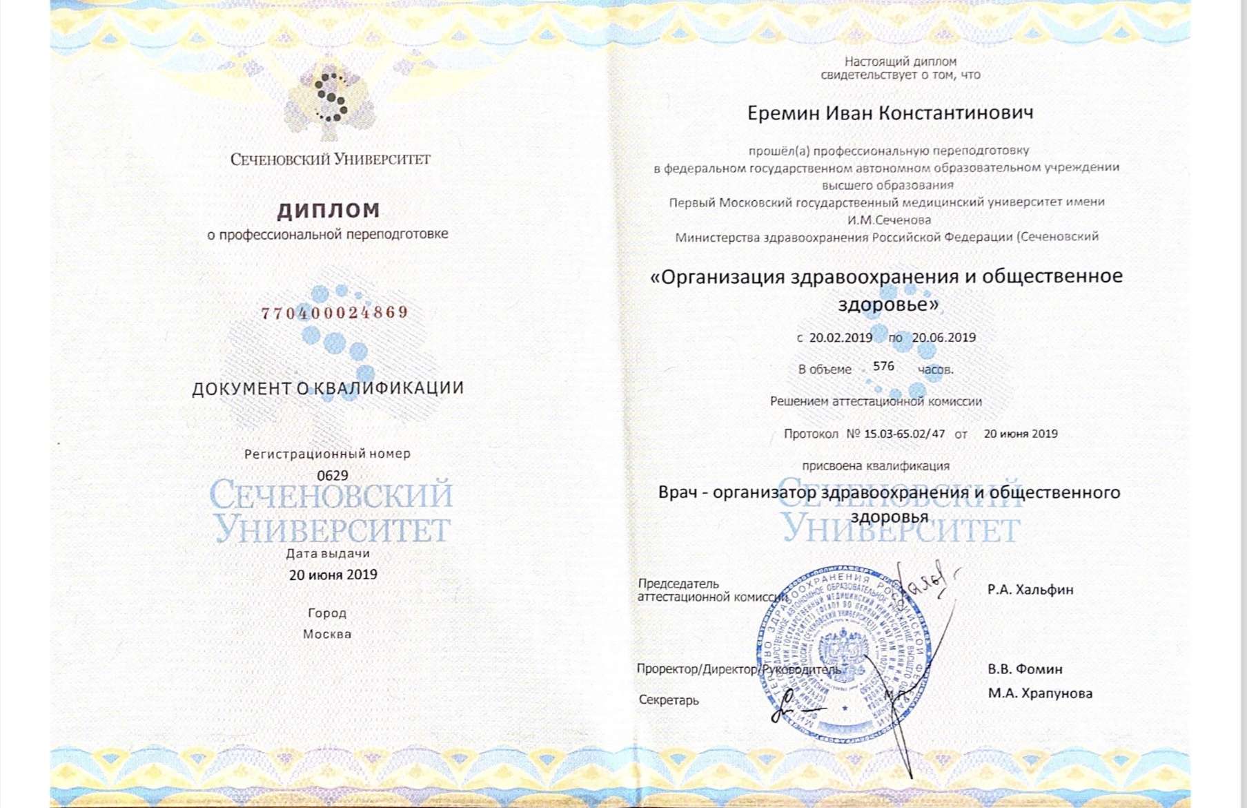
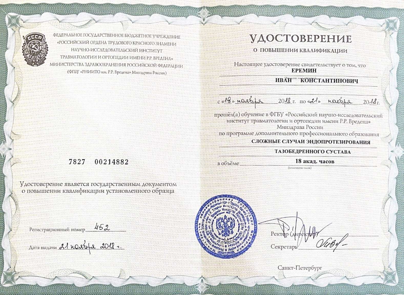
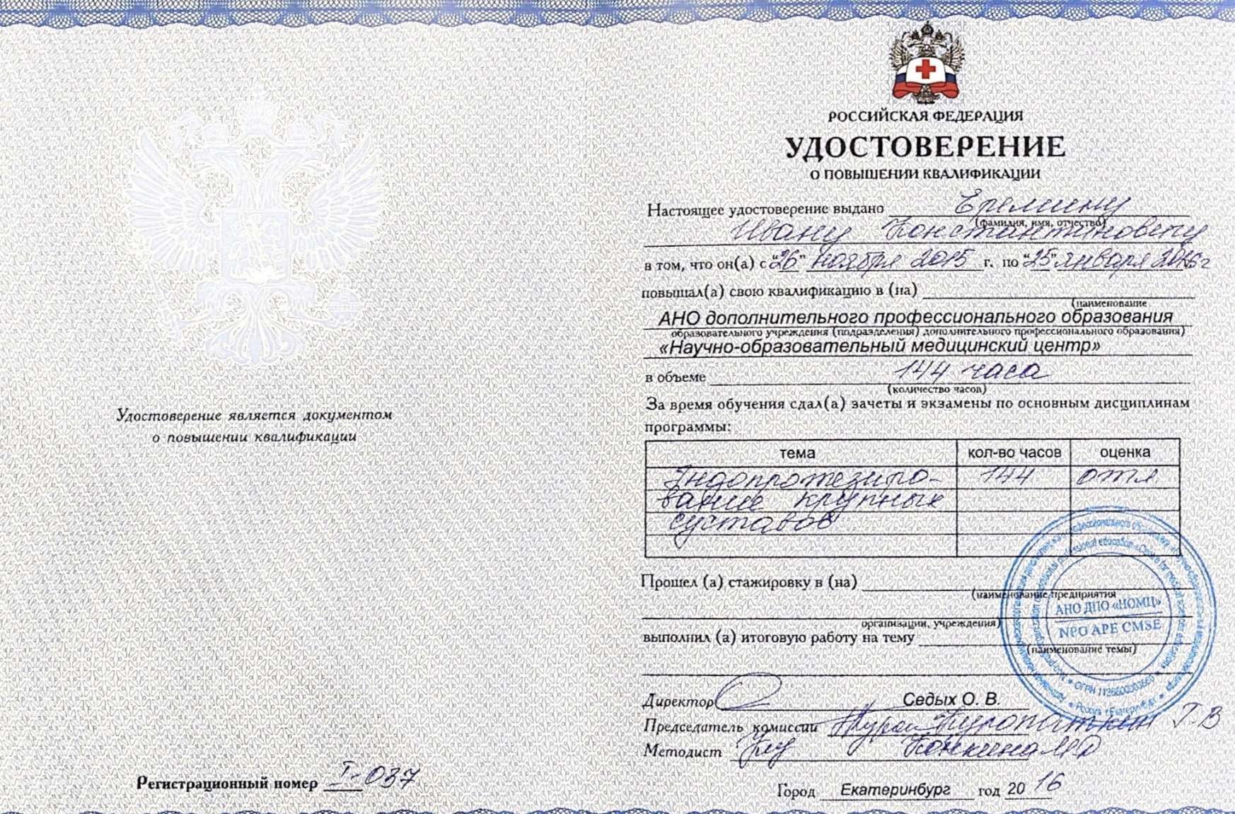
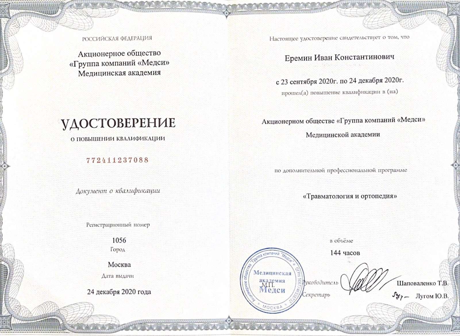
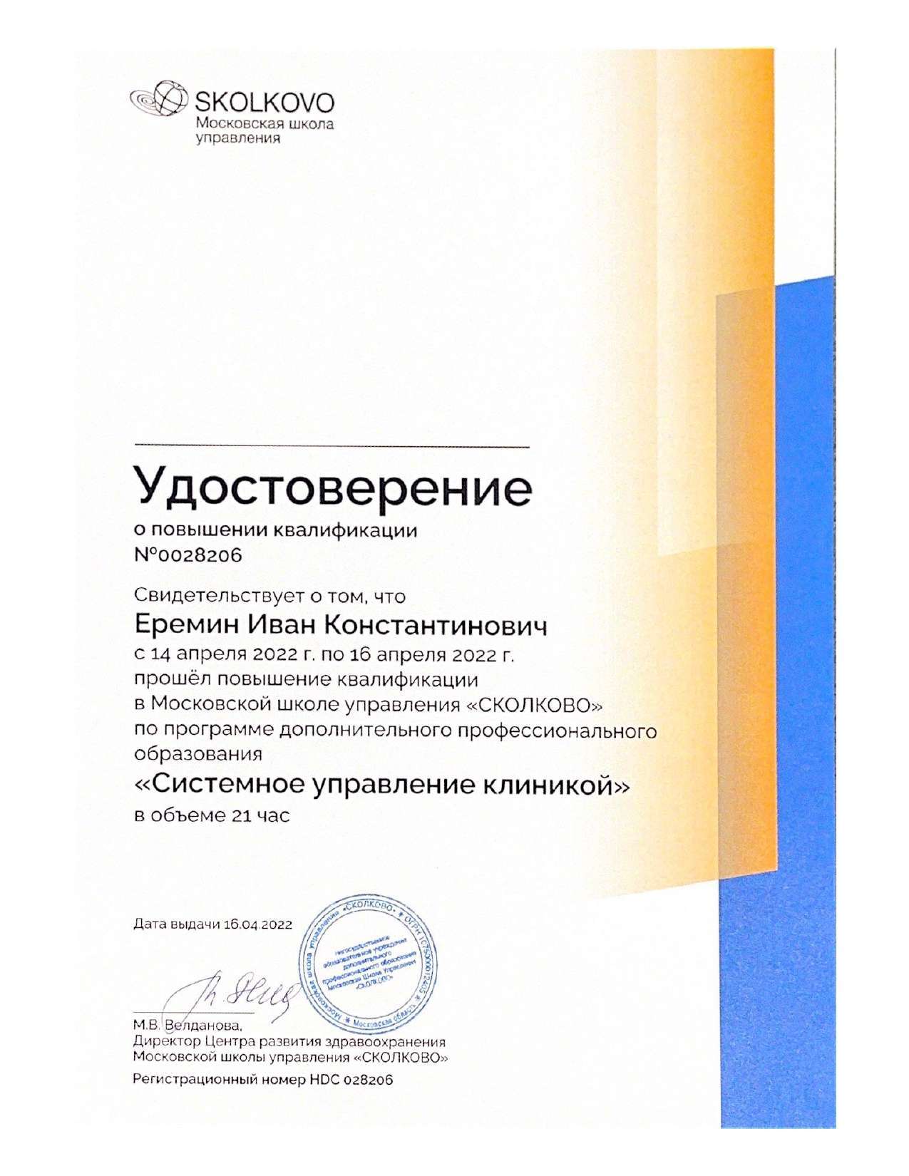
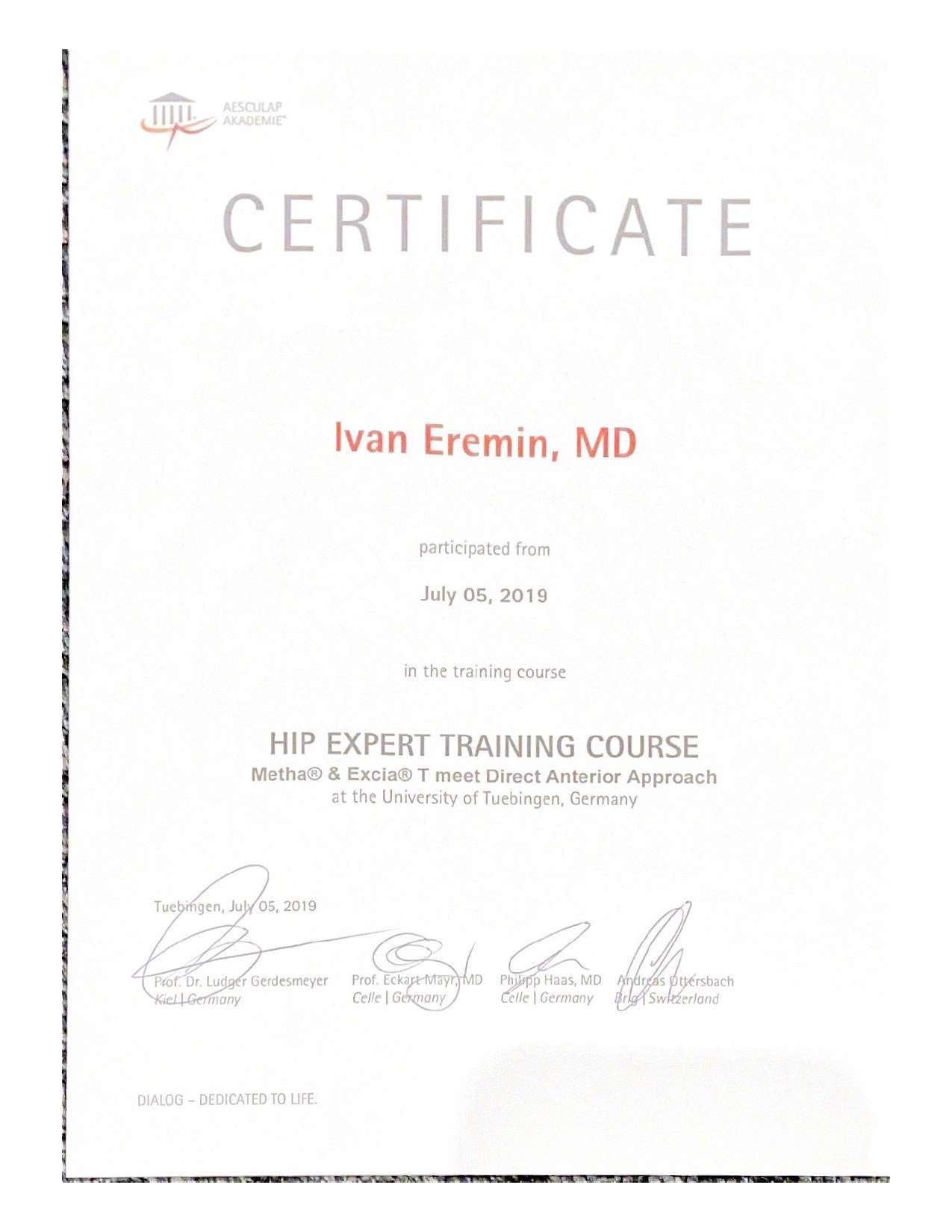
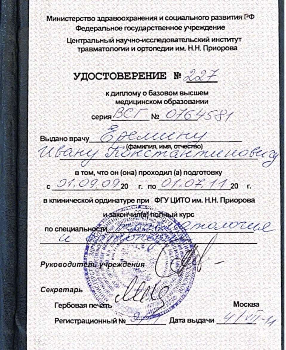
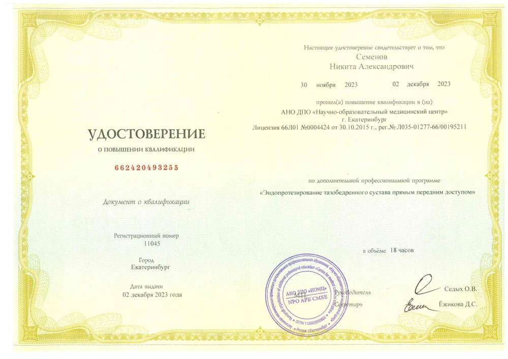
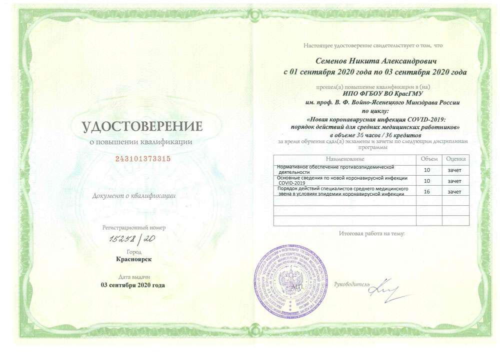
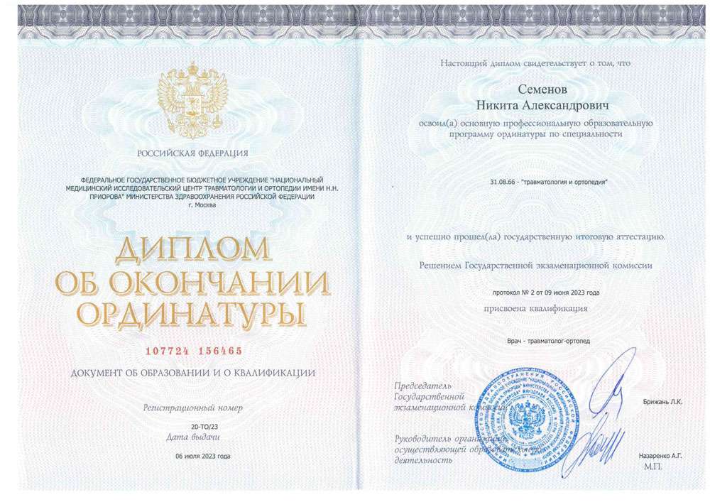
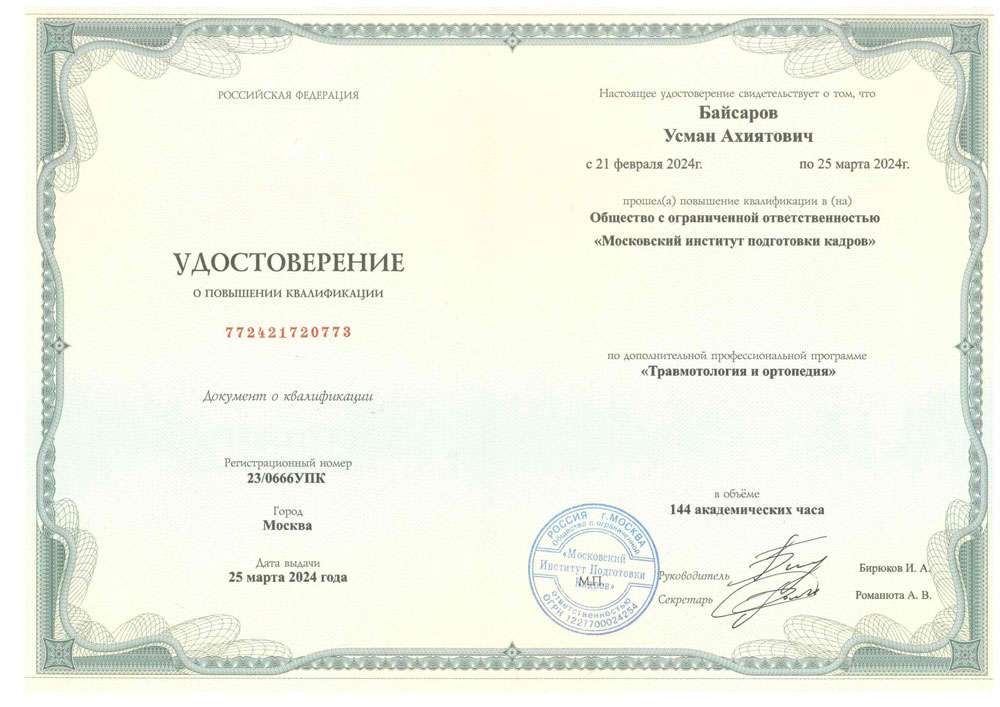
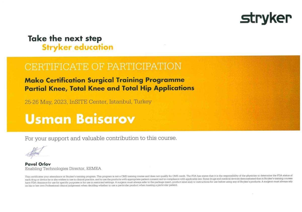
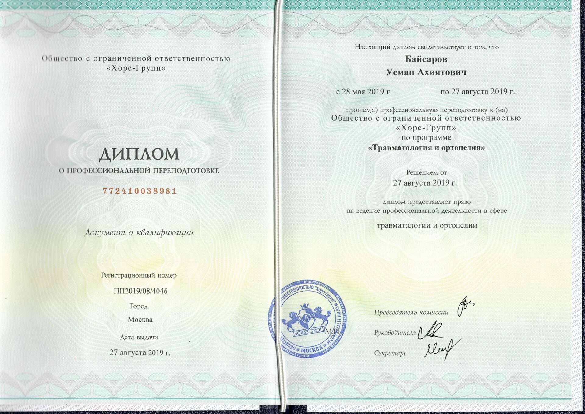
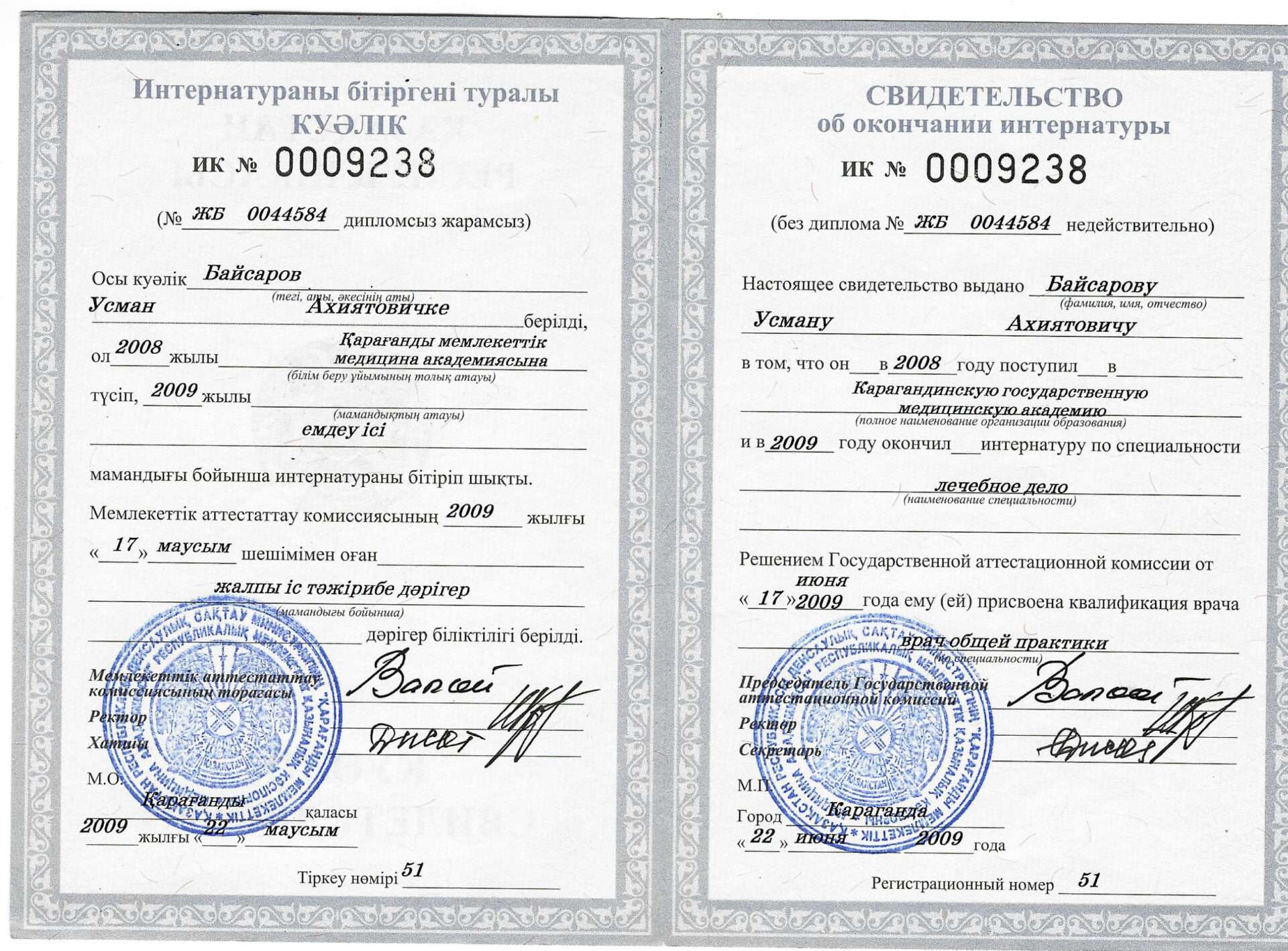
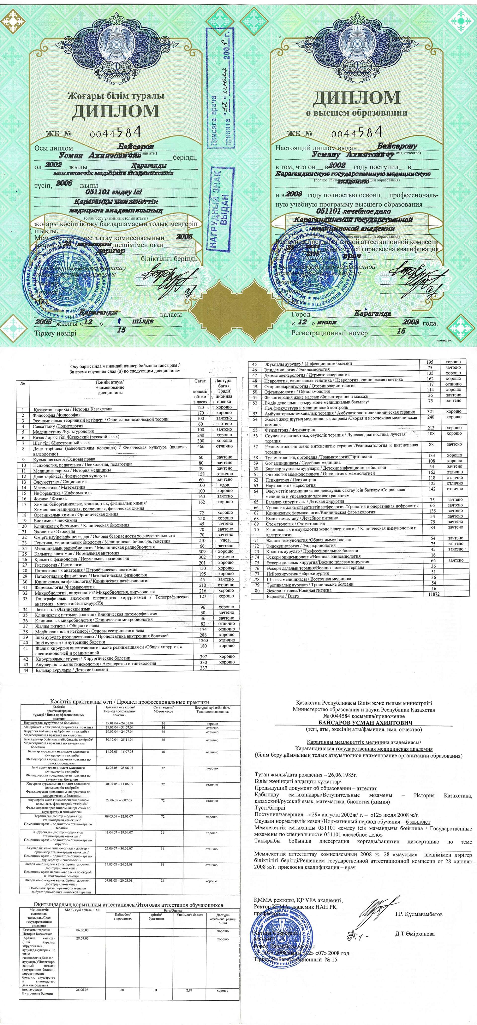
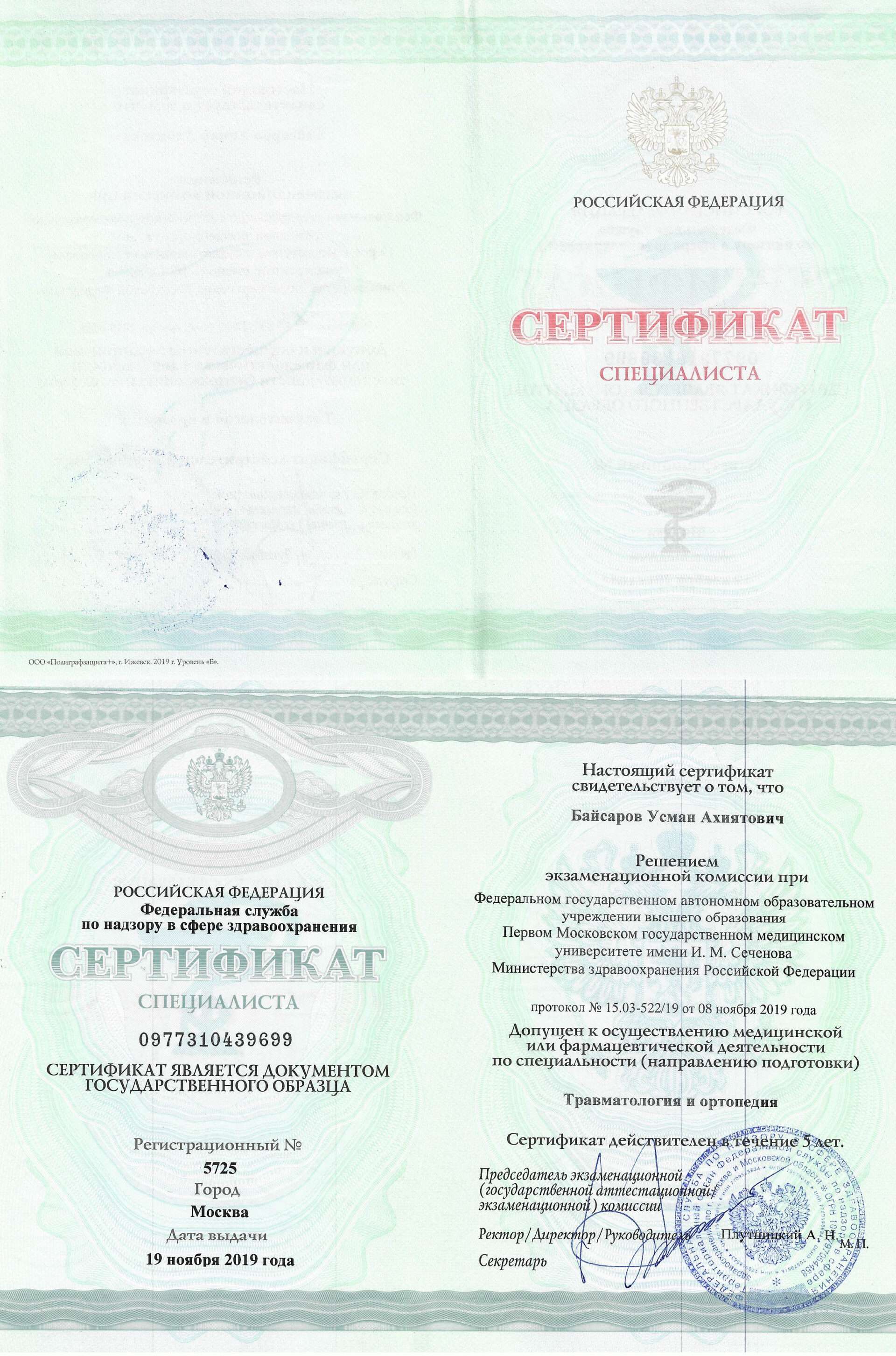
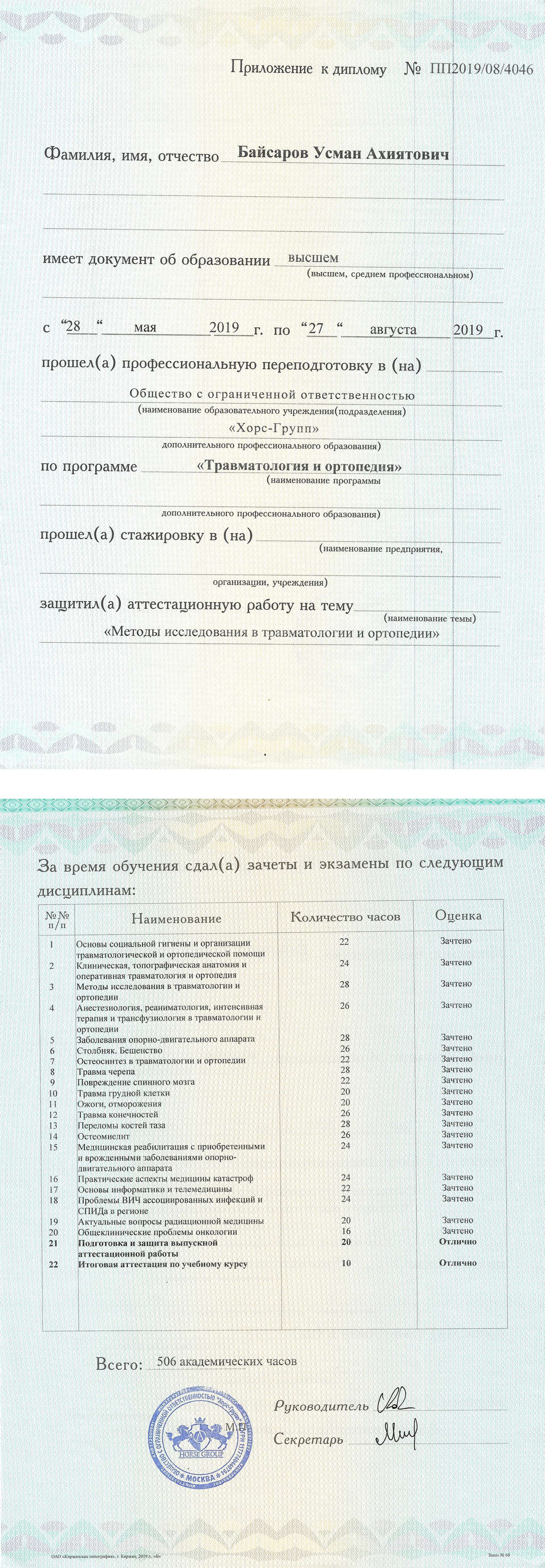
Сайт может отображаться некорректно, поскольку вы просматриваете его с устаревшего браузера Internet Explorer (), который больше не поддерживается Microsoft.
Рекомендуем обновить браузер на любой из современных: Google Chrome, Яндекс.Браузер, Mozilla FireFox.
Пожалуйста, поверните устройство в вертикальное положение для корректного отображения сайта

Femoral neck fracture osteosynthesis

  • High success rate:
    Over 90% of patients report significant improvement after hip treatment.
  • Individual approach:
    Personalized treatment programs for each patient.
  • Modern technology:
    Using the latest medical advances for maximum effect.

Hip fracture

This is an anatomical impairment of the integrity of the femur, which practically does not respond to conservative treatment. In most cases, the pathology is treated by surgery, one of the types of which is osteosynthesis. Osteosynthesis in a hip fracture is an operation in which the reduction of fragments of the femur and their rigid fixation are performed.

Purpose of osteosynthesis

The main goal of the hip fracture operation is to recreate the correct position of bone fragments in the fracture zone, maintaining their immobility until the final fusion of the femoral neck.

During hip surgery, various designs are used — metal spokes, threaded and rod systems (screws), devices with diaphyseal plates. Systems for osteosynthesis are made of special alloys of metal based on titanium or steel, which must be resistant to corrosion and biocompatible with the tissues of the human body.

Indications and contraindications for femoral osteosynthesis

Surgery of reposition (match in the correct position) of fragments in case of hip fracture is required in the following cases:

  • Displaced fracture. Displaced fracture.
  • Comminuted fracture. Comminuted fracture.
  • Combined injury.
For example, a fracture combined with a dislocation. Combined injury.
    For example, a fracture combined with a dislocation.
  • Improper bone fusion, pseudoarthrosis, etc. Improper bone fusion, pseudoarthrosis, etc.
h
  • decompensated diseases of various organs and systems;
  • severe general condition of the patient, shock, coma;
  • infectious diseases, active form of tuberculosis;
    coagulopathies;
  • purulent inflammations, skin, soft tissue, bone infection in the intervention area;
  • severe mental impairment;
  • severe lower extremity thrombophlebitis.

For a successful result of osteosynthesis in case of a hip fracture, its location, the degree of impaired blood supply to the bone in the injury area is also taken into account. Subcapital fracture with displacement, significant disruption of blood supply to the femoral head, the likelihood of aseptic necrosis significantly reduce the likelihood of successful intervention.

Methods of osteosynthesis

Osteosynthesis in hip fracture is performed by two main methods — extramedullary and intramedullary. Both of these methods are a type of internal (immersion) osteosynthesis, however, with the extramedullary method, the structural elements do not affect the medullary canal, and with the intramedullary one, they are passed through it.

Intramedullary method

Intramedullary osteosynthesis is one of the most effective treatments for fractures. It involves the introduction of special devices into the medullary canal of each of the bone fragments. These devices are rods, nails, which can be rigid or semi-elastic.

To ensure the stability of the system, there are screw holes or special bends on the distal ends of the fixing elements. Structures for intramedullary osteosynthesis can even fix fractures with many fragments.

The nails are inserted into the medullary canal after pre expansion or without it. After bone fusion, all intramedullary constructs are removed.

Extramedullary osteosynthesis

For osteosynthesis with screws in case of hip fracture, doctors use special plates, which are attached to the bone with fasteners.

Preparation for hip
osteosynthesis surgery

In case of a fracture of the femoral neck, surgery is recommended to be carried out as quickly as possible, so preparation for it should take place in an accelerated mode.

Confirmation of diagnosis

To confirm the diagnosis of hip fracture, and to determine its nature, radiography is carried out. Computer or magnetic resonance imaging will help to assess the degree of impaired blood supply to the fracture area, which affects the possibility of osteosynthesis.

Standard preparation

Standard preparation includes cleansing the intestines with an enema or special drugs the night before and on the morning of the operation, removing hair in the surgical area. The last meal (light dinner) should be 8-10 hours before the intervention, it is done on an empty stomach, you cannot eat or drink on the morning of the operation.


If there are concomitant pathologies

It is necessary to obtain the medical conclusion of the attending physician about the possibility of surgery.

On the previous night or immediately before the operation, the patient talks with an anesthesiologist, who will find out whether surgical treatment was carried out earlier, what type of anesthesia was used, whether the patient is allergic to drugs, whether he abuses alcohol, what medicines he is on.

This data will help the doctor choose the optimal anesthetic support. In most cases, osteosynthesis for femoral neck fracture is performed under general anesthesia. In some cases, epidural anesthesia is possible.

Additional examination

The patient is also prescribed an additional examination:

  • complete blood count and urine tests;
  • biochemical blood test, determination of blood type and Rh factor;
  • hemostasiogram;
  • a test to determine indicators of mineral metabolism (phosphorus, calcium);
  • densitometry;
  • ECG;
  • Photofluorography or chest X-ray.
Mandatory list of tests
  1. Detailed (clinical) complete blood count with platelet count, up to 14 days.
  2. Urinalysis, not more than 14 days.
  3. Biochemical blood test (ALT, AST, total and direct, bilirubin, blood glucose, total protein, urea, creatinine, sodium, calcium, potassium, chlorine), for no more than 14 days.
  4. Coagulogram (fibrinogen, APTT, prothrombin time, Quinque prothrombin time, INR, thrombin time, clotting time and bleeding duration), up to 14 days.
  5. Blood test on hepatitis markers In (HbsAg) and hepatitis C (HCV), no more than 3 months.
  6. Blood test on RW, up to 1 month.
  7. Blood test on HIV infection, no more than 3 months.
  8. Blood type, Rhesus factor, up to 2 months.
  9. Phenotyping of erythrocytes on Rh system antigens (C, E, c, e) and Kell antigen, it is termless
  10. Alloimmunny anti-erythrocyte antibodies, including antibodies to Rh-antigen
  11. The electrocardiogram (ECG) with interpretation and the conclusion, up to 14 days.
  12. Fluorographic examination of the chest organs or a chest X-ray in 2 projections, for a period not exceeding 6 months.
  13. Fibrogastroduodenoscopy, not more than 3 months. In the presence of ulcerative history, erosion of the gastrointestinal tract, not more than 1 month.
  14. Ultrasound examination of the veins of the lower extremities, up to 1 month.
  15. Ultrasound examination of the arteries of the lower extremities, for a period not exceeding 1 month.
  16. Ultrasound examination of brachiocephalic arteries, not more than 3 months.
  17. Echocardiography (cardiac ultrasound), not more than 1 month.
  18. Daily ECG monitoring, for individuals with hypertension over 65 years of age, not more than 1 month.
  19. TSH, glycated hemoglobin, for individuals with diabetes mellitus, not more than 14 days.
  20. Conclusion from the therapist in printed form, indicating the absence of contraindications to surgical treatment and indicated concomitant diseases, up to 1 month
  21. Conclusion from narrow specialists (oncologist, endocrinologist, cardiologist, etc.) complete examination, in the presence of concomitant diseases, not more than 1 month
  22. X-ray of pelvic bones with capture of the upper third of the thigh, femur in one projection on the film at 100% scale. (In one image, both joints are in direct projection).

Center staff

Ivan Konstantinovich Eremin
The main surgeon
Nikita Alexandrovich Semenov
Orthopedic traumatologist
Usman Akhiyatovich Baysarov
Orthopedic traumatologist
Zurab Malkhazievich Molarishvili
Orthopedic traumatologist
Svetlana Vladimirovna Fedichkina
Operating nurse in traumatology and orthopedics
Ekaterina Vasilievna Dolzhikova
Administrator
Anna Mikhailovna Yazykova
Administrator
Ivan Konstantinovich Eremin
The main surgeon

Work experience 15 years

Specialization:
-Hip arthroplasty via the DAA
-Total hip and joint arthroplasty using a robot assistant MAKO
-Revision hip surgery

Certificates
Nikita Alexandrovich Semenov
Orthopedic traumatologist

Work experience 3 years

Specialization:
-Hip arthroplasty via the DAA
-Revision hip surgery
-Conservative treatment of hip joint

Certificates
Usman Akhiyatovich Baysarov
Orthopedic traumatologist

Work experience 13 years

Doctor of the highest category

Specialization:
-Hip arthroplasty via the DAA
-Total hip arthroplasty with robot assistant MAKO
-Revision hip surgery
-Conservative treatment of hip joint

Certificates
Zurab Malkhazievich Molarishvili
Orthopedic traumatologist

Work experience 5 years

Specialization:

-Hip arthroplasty via the DAA
-Total hip arthroplasty with robot assistant MAKO
-Revision hip surgery
-Conservative treatment of hip joint

Certificates
Svetlana Vladimirovna Fedichkina
Operating nurse in traumatology and orthopedics

Work experience 15 years

Certificates
Ekaterina Vasilievna Dolzhikova
Administrator
Certificates
Anna Mikhailovna Yazykova
Administrator
Certificates

Operation progress

01

The patient is placed on the operating table and put into anesthesia. During the operation, they will not feel anything and will come to their senses in the intensive care unit, where they will be under close supervision of medical personnel.

02

The limb is fixed in the desired position. The operating field is treated with an antiseptic. Then the surgeon makes an incision, the size of which is from 2-3 to 10-15 cm, depending on whether a closed or open reposition is carried out.

03

In closed intramedullary osteosynthesis, the fixation construct is inserted at a specific point in the femoral head under radiographic control through a 2-3 cm incision by screwing in and using special instruments.

04

Open osteosynthesis involves an extensive incision (up to 10-15 cm), comparison of bone fragments, installation of the selected fixation system.

05

The final stage of the operation is the treatment of the wound, its drainage, and suturing. If the chosen technique involves immobilization of the limb, a plaster cast is applied to the patient.

.

At the Hip Surgery Center, osteosynthesis for hip fracture is carried out using modern methods and high-tech equipment. Experienced doctors of the clinic are constantly improving their qualifications, using only the best experience in their activities.

Employees of the Center make every effort to ensure that the treatment process, recovery of patients proceed as quickly as possible, comfortably and with the best results.

Rehabilitation

Immediately after the operation, the patient is transferred to the intensive care unit or ward, a cold compress is applied to the place of intervention, antibiotic therapy, painkillers, and, if necessary, other drugs are prescribed. Bed rest after surgery should not be long, maximum 1-2 days, early activation contributes to the prevention of postoperative complications, faster recovery. However, the load on the operated limb should be reduced, the patient should move using crutches. In the early recovery period, breathing exercises and massage are carried out to prevent congestion. It is necessary to treat the sutures, which are removed in 10-12 days.

After the operation wound is healed, the patient is prescribed recovery procedures, the number and intensity of which increases gradually. These include:

  • physiotherapy exercises — passive and active exercises, exercises on simulators;
  • physical therapy;
  • therapeutic baths;
  • paraffin wraps;
  • massage;
  • swimming, aquagymnastics.

The time to fully restore the functions of the limb and return to the usual way of life depends on many factors: the patient’s age, state of health and physical shape in general, the severity of the fracture, the absence or presence of postoperative complications. The rehabilitation period can take from 3-6 to 12 months.

Don’t let joint pain define your life

Give yourself freedom of movement without restrictions

Over 5 years, using the DAA method, we returned more than 3 thousand patients to an active life.

Submit a request and get a consultation